Can You Get a Gum Graft on Dental Implants or Veneers?
If you are researching options in New York, start with our page on gum disease and gum grafting treatment in Brooklyn.
Table of Contents
1. Introduction
When gums recede around natural teeth, the exposed root surfaces can cause sensitivity, higher risk of decay on the root, aesthetic concerns, and in some situations a pathway for further periodontal breakdown. Gum grafting, also called soft tissue grafting or periodontal plastic surgery, is widely used to rebuild lost gum tissue and improve both health and appearance.
Many patients ask a more complex version of this question. What if the tooth is restored with a veneer or a crown. What if the tooth was previously extracted and replaced with a dental implant. Can gum grafting still be done? The short answer is that gum grafting can be very effective around implants and sometimes around veneered or crowned teeth. There are important caveats. The biology around implants is different from that of natural teeth, veneer margins can complicate healing, and predictable results require careful planning. This article explains the science and the clinical decision making so you can understand what is possible, what is not, and what steps help create a predictable outcome.
2. Basic anatomy and biology: gums, implants, veneers
To understand what is feasible, it helps to review how soft tissue attaches and heals in different settings.
Natural teeth have a periodontal ligament that connects the root to bone. Collagen fibers insert into the root cementum, and there is a junctional epithelium at the sulcus. Blood supply comes from multiple sources. When gums recede, cementum and sometimes dentin are exposed.
Dental implants are titanium or similarly biocompatible fixtures placed in bone. They do not have a periodontal ligament. Around implants, soft tissue fibers tend to orient parallel to the implant or abutment instead of inserting into it. Vascular supply is different and more limited than around a natural tooth. The width and thickness of keratinized mucosa around the implant matter for long term stability. Thin soft tissue phenotypes are associated with a greater risk of soft tissue recession and inflammation. A recent review on implant outcomes and phenotype summarizes these relationships in detail and highlights the importance of soft tissue thickness and quality around implants (implant outcomes and gingival phenotype review).
Veneers are thin porcelain or composite shells bonded to the front of teeth. Veneers can have margins near or slightly under the gumline. Veneers change the contours and the cleanability of the tooth. Veneers do not regenerate gum tissue. If margins are not ideal or if hygiene is difficult, chronic inflammation can develop and recession can progress.
Because grafts rely on a well vascularized recipient bed and a biologic attachment to natural tissues, these differences in anatomy influence what techniques will work and how predictable they are.
3. Why gum recession occurs around implants and veneers
Recession near implants or veneered teeth can happen for many reasons that overlap with natural teeth, along with restoration specific issues.
Traumatic toothbrushing or oral habits that apply repetitive mechanical stress
Inflammation from plaque accumulation when margins are difficult to clean
Ill fitting or overcontoured veneer or crown margins that create plaque traps
Thin tissue phenotype that is more susceptible to shrinkage
Low width of keratinized mucosa around implants
Surgical factors at implant placement such as tissue thickness, flap management, and implant position
Underlying crestal bone remodeling or bone loss
Smoking and systemic risk factors that impair healing
Before a graft is considered, the cause or combination of causes should be identified and addressed. Otherwise the same factors that caused recession can undermine graft success.
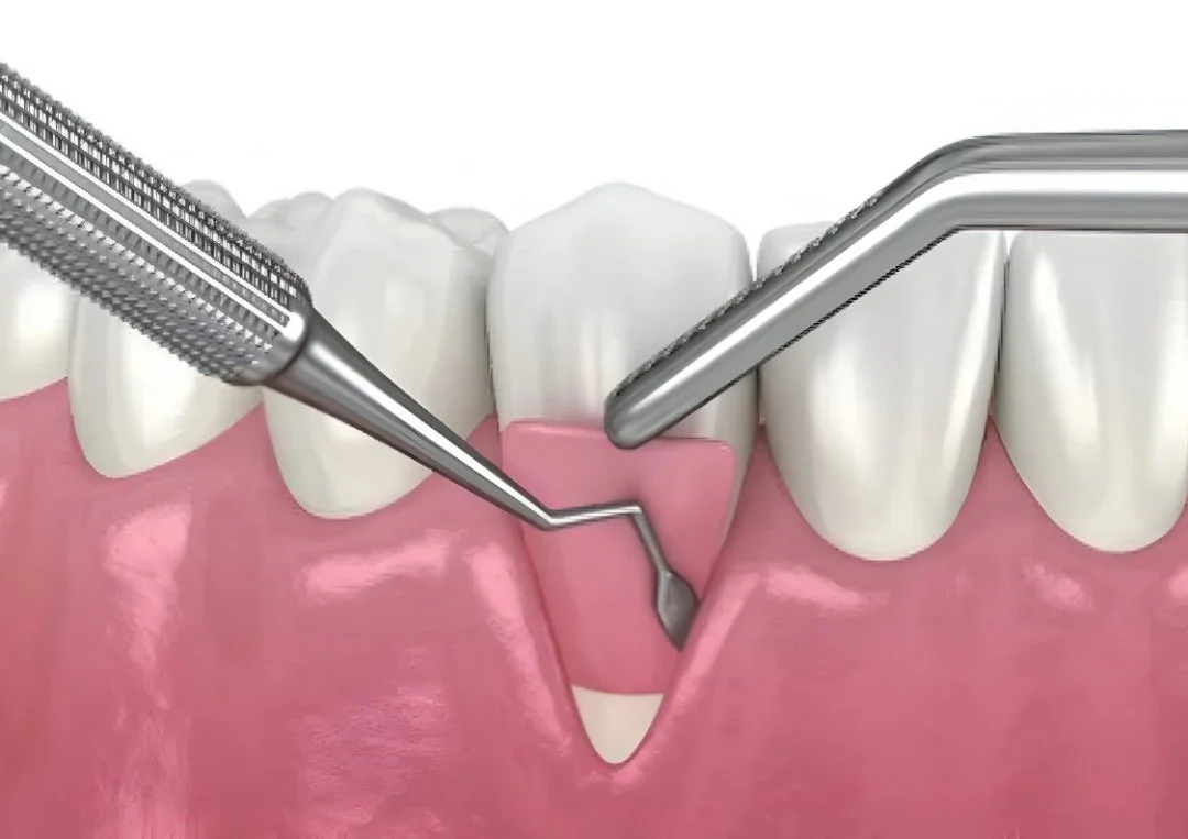
4. What a gum graft is: types and purpose
Primary goals of gum grafting
Increase the thickness of soft tissue for durability and improved seal
Increase the width of keratinized tissue around implants and teeth
Cover exposed roots to reduce sensitivity and risk of root caries
Improve aesthetic symmetry of the gum line and smile
Create a more stable soft tissue barrier that resists bacterial challenge
Common graft types
Subepithelial connective tissue graft, CTG. Autogenous connective tissue is harvested from the palate and placed under a flap to thicken and stabilize the recipient site.
Free gingival graft, FGG. A thin, epithelialized graft from the palate is transplanted to increase keratinized tissue width, often used in the posterior or around implants where root coverage is not the primary goal.
Pedicle flaps. Tissue adjacent to the recession is repositioned to cover the defect.
Soft tissue substitutes such as xenogeneic collagen matrices or acellular dermal matrix. These reduce donor site morbidity and can be effective when thickness requirements are modest or when a patient prefers to avoid a palatal harvest.
Each material and technique has trade offs related to patient comfort, color match, thickness gained, and long term dimensional stability.
5. Scientific evidence for grafts around implants
There is substantial and growing literature on soft tissue augmentation adjacent to dental implants.
A controlled comparison of palatal connective tissue grafts versus a xenogenic soft tissue substitute for augmentation around implants found that CTG tends to achieve greater soft tissue thickness and stability, although substitutes can reduce morbidity and still deliver clinically useful improvements in many cases. See the full study on the National Library of Medicine site: soft tissue augmentation around dental implants with CTG versus xenogenic substitute.
Soft tissue grafting in the management of peri implantitis has been investigated as an adjunctive therapy to improve soft tissue conditions and facilitate hygiene after decontamination. Clinical reports support that grafting can improve peri implant soft tissue health in selected cases. Read the open access article here: soft tissue grafting for peri implantitis.
A prospective effort is comparing free gingival grafts and other methods for maintaining peri implant mucosa morphology, which underscores interest in the best way to develop durable keratinized tissue around implants. See the registered protocol: peri implant mucosa morphology trial NCT06065254.
Clinical observations comparing tooth and implant sites suggest that free gingival grafts effectively increase keratinized tissue width around implants, with some differences in dimensional change compared to natural teeth. See the journal report: clinical observations of soft tissue dimensions around tooth and implant sites.
A recent narrative and systematic review on gingival phenotype and implant outcomes highlights that thin tissue phenotypes correlate with greater risk of soft tissue recession and marginal bone changes, supporting the rationale for soft tissue thickening in at risk cases. Read more here: implant outcomes and gingival phenotype review.
Clinical takeaway. Around implants, soft tissue augmentation is a mainstream, evidence supported strategy to strengthen the mucosal seal, increase keratinized tissue width, and improve aesthetic contours. Autogenous CTG remains a reference standard when maximum thickness and stability are required. Substitutes are useful for selected indications and to reduce donor site morbidity.
6. Scientific evidence in the presence of veneers or crowns
The literature that focuses narrowly on grafting near existing porcelain veneer margins is more limited than the implant literature, but several themes recur.
When recession is severe and the tissue is thin, a restorative approach can sometimes be chosen instead of or in combination with surgery, especially in the esthetic zone. An example is a case report describing the restoration of recession with preformed periodontal composite veneers when surgical root coverage was not predictable. See: restoration of recession with periodontal preformed composite veneers.
Many interdisciplinary esthetic reports recommend grafting before definitive veneers or crowns so that the final restoration margins can be designed in harmony with the healed soft tissues. A recent review illustrates planning and sequencing principles: soft tissue grafting procedures before restorations in the esthetic zone.
Experienced clinicians consistently caution that soft tissue grafts cannot biologically attach to porcelain the way they do to natural root cementum. If a porcelain or metal margin is the only recipient surface, true connective tissue attachment is not expected. The clinical aim becomes thickening and contour improvement rather than stable coverage over the porcelain. For a patient facing explanation, see this Q and A: gum grafts with existing veneers or crowns.
Patient education resources also emphasize that veneers themselves do not treat gum loss and that surgical soft tissue augmentation or margin redesign may be required for long term stability and easy hygiene. Example overview: porcelain veneers and gum recession overview.
Clinical takeaway. Around veneers and crowns, grafting is most predictable when there is accessible natural tooth structure adjacent to the margin, when the margin can be modified or ideally redesigned after graft healing, and when inflammation is fully controlled.
7. Can you graft over veneer material or porcelain
This is the heart of the question. In general, you cannot achieve a classic connective tissue attachment over porcelain or composite. However, grafting can still help in several ways if the clinical conditions are right.
Why a graft does not work directly over porcelain
Attachment biology. Connective tissue fibers insert into root cementum and align with natural tooth structures. On porcelain or metal, the tissue heals with an epithelial adaptation at best rather than a robust connective tissue insertion.
Margin related inflammation. Subgingival or overhanging margins accumulate plaque and can create chronic inflammation that prevents graft integration.
Recipient bed vascularity. A successful graft needs a well vascularized bed. A smooth, inert material does not provide the vascular support that living tissues provide.
Bone level and biologic width. If the bone crest is too close to the margin or if the biologic width has been violated, the tissue will remodel and pull away, limiting stable coverage.
When grafting still helps near veneers
If part of the root is exposed adjacent to a veneer, a CTG with a coronally advanced flap can thicken the tissue and partially cover the root, which improves sensitivity and aesthetics even if the edge of the veneer remains at or near the margin.
If veneers will be replaced after healing, grafting first allows the final margin to be placed in a position and contour that the tissue can support, which improves long term stability.
In cases with minimal recession but a very thin biotype, a thicken and stabilize approach can reduce the risk of future recession without attempting full coverage over porcelain.
8. Clinical feasibility: when it can work, when it cannot
| Factor | Favorable for grafting | Unfavorable for grafting |
|---|---|---|
| Recipient surface | Natural root or accessible implant soft tissue with adequate keratinized mucosa | Purely porcelain or metal margin with no natural root exposure |
| Restoration margin | Supragingival or shallow, well polished, hygienic | Deep subgingival, overhanging, or rough margin that traps plaque |
| Tissue phenotype | Thick or moderate, capable of supporting a flap and graft | Thin biotype with minimal keratinized tissue and high contraction risk |
| Bone support | Stable crestal bone and intact biologic width | Significant bone loss or biologic width violation |
| Patient factors | Excellent hygiene, non smoker, stable systemic health, good compliance | Poor hygiene, smoking, uncontrolled systemic conditions |
| Sequencing | Ability to graft first then place or replace veneers or crowns | Veneers cannot be modified or replaced and margins are not acceptable |
If most favorable boxes are checked, grafting can succeed and provide stable improvement. If most unfavorable boxes are present, a restorative or prosthetic approach may be more predictable.
9. Treatment planning: steps, preconditions, patient factors
Comprehensive assessment
Record periodontal charting, recession depth, keratinized tissue width, probing depths, phenotype, and photograph the smile line. Take radiographs and, for implants, consider a CBCT if tissue deficiency may be related to implant position.Etiology control
Eliminate inflammation before surgery. Polish or temporarily modify overcontoured margins. Provide hygiene coaching and tools such as interdental brushes and water flossers. Address traumatic brushing.Define patient goals
Clarify whether the primary goal is sensitivity relief, improved aesthetics, durable thickness for long term health, or all of the above. Align expectations around how much coverage is realistic when veneers or implants are present.Sequence the interdisciplinary care
Whenever possible, plan grafting before definitive veneer or crown placement. For existing restorations, discuss temporary removal to improve access and recipient bed quality, then replace after healing.Choose the graft type
CTG is preferred when maximum thickness and long term stability are desired, especially around implants where thickness is protective. See comparative data here: CTG versus xenogenic soft tissue substitute around implants.
FGG is useful to increase keratinized tissue width around implants where the goal is a more durable, hygienic collar rather than root coverage. See clinical comparisons here: soft tissue dimensions at tooth and implant sites with FGG.
Soft tissue substitutes can be considered to reduce palatal morbidity in selected cases, with the understanding that dimensional stability may be less than CTG.
Plan post operative maintenance
Schedule follow ups to monitor healing, reinforce hygiene, and coordinate restorative steps. For implants, ensure the final restoration has a cleanable emergence profile and polished transmucosal surfaces.
10. Surgical techniques and materials for implants and veneers
Around implants
Connective tissue graft with split thickness flap. This is a common approach for thickening peri implant mucosa and improving the emergence profile. The graft is stabilized around the abutment or provisional crown with sutures.
Free gingival graft for keratinized tissue width. Especially in posterior regions, an FGG can create a stable band of keratinized mucosa that improves comfort and hygiene.
Substitutes may be used as a collagen matrix placed under a partial thickness flap. These are helpful when a palatal harvest is undesirable. Evidence supports their use, though the magnitude of thickness gain may be less than CTG. See the controlled comparison here: CTG versus xenogenic soft tissue substitute around implants.
Near veneers and crowns
Coronally advanced flap with CTG when a portion of root is exposed. The root surface is planed and conditioned as indicated, and the tissue is advanced to partially cover the exposure. The veneer margin may be polished or redesigned later.
Margin modification or provisionalization. If a margin is subgingival and overcontoured, provisional restorations with better contour can be placed during healing to reduce plaque retention and allow the graft to mature.
Interproximal papilla preservation and gentle flap handling reduce recession at the embrasures and help maintain papilla height in the esthetic zone.
Peri implantitis scenarios
When soft tissue is thin and inflamed, grafting may be combined with decontamination and regenerative or resective procedures to create a more maintainable environment. See clinical background here:soft tissue grafting for peri implantitis.
11. Healing, outcomes, expected results, limitations
Timeline
First 7 to 14 days. Initial epithelialization, management of swelling, and suture removal as indicated.
Weeks 3 to 6. Tissue maturation and early contraction. Patients should protect the area and avoid trauma.
Months 3 to 12. Final contour and color continue to refine. Minor shrinkage can occur over time, especially with thin phenotypes or substitute materials.
Aesthetic expectations
Around implants, the main goals are a thicker and more stable collar of tissue and an emergence profile that looks natural when smiling.
Around veneers, full coverage over porcelain margins is rarely realistic. Expect improvement in thickness, contour, and sometimes partial root coverage, with final aesthetics optimized by carefully designed restoration margins after healing.
Functional outcomes
Thicker tissue can reduce sensitivity and improve patient comfort.
A wider band of keratinized mucosa around implants often improves hygiene and reduces mucosal inflammation.
Limitations
Grafting over a purely porcelain surface does not create a true connective tissue attachment.
Severe bone deficiencies limit the coronal position that tissue can maintain.
Thin tissue phenotypes are more prone to contraction, which can modestly reduce the initial coronal gain.
12. Risks, complications, and how to manage them
Graft partial loss or necrosis. Minimize by ensuring a tension free flap, firm stabilization, and adequate blood supply.
Color or texture mismatch. CTG often provides superior blending. Substitutes may differ slightly in appearance.
Persistent inflammation from margins. Manage by adjusting or replacing restorations with polished, hygienic profiles.
Donor site discomfort. Discuss analgesia, palatal dressing, and dietary modifications. Substitutes can reduce this risk.
Recession relapse. Reinforce gentle hygiene, use ultra soft brushes, and monitor for traumatic habits such as nail biting or aggressive flossing.
13. Case studies and practical examples
Implant tissue thickening to stabilize the collar. A posterior mandibular implant with minimal keratinized mucosa and tissue thickness is grafted with CTG to create a thicker, more resilient band of tissue. This reduces bleeding on probing and improves comfort on brushing. The comparative data supporting CTG for thickness gain can be reviewed here: CTG versus xenogenic soft tissue substitute around implants.
Esthetic zone veneer case with prior recession. A patient presents with thin tissue and recession on a maxillary lateral incisor that has an aging veneer. A coronally advanced flap with CTG is performed to thicken and partially cover the root. After three months, the veneer is replaced with a margin designed to be supragingival where possible and smoothly polished where proximity to tissue is required. This sequence mirrors recommendations discussed in this review: soft tissue grafting procedures before restorations.
Peri implant mucositis that progresses to peri implantitis. After decontamination and correction of an overcontoured crown, a soft tissue graft is added to increase thickness and facilitate hygiene. Background and rationale appear in this review:soft tissue grafting for peri implantitis.
14. FAQs
-
Not in a biologic sense that creates a connective tissue insertion. Grafting can still improve thickness and contour near the veneer, but true coverage over porcelain with stable attachment is not predictable.
-
Sometimes. If the margin is deep, overcontoured, or inflamed, provisionalization or replacement is often advised so the recipient bed is healthy and accessible. Many interdisciplinary teams plan grafting first and definitive veneers second.
-
Yes, if exposed root is present next to the veneer, a graft can reduce sensitivity by covering or thickening over the root. Veneers themselves do not treat root sensitivity if the exposure is apical to the veneer margin.
-
Initial healing takes one to two weeks, with full maturation over several months. Your periodontist will instruct you on a protected hygiene routine. Typically, the site is not brushed for the first one to two weeks, and then gentle brushing with an ultra soft brush is introduced.
-
Substitutes can work well in selected cases and reduce donor site discomfort. When maximum thickness and long term stability are required, CTG still shows advantages in controlled comparisons. See the study here: implant soft tissue augmentation with CTG versus substitute.
-
A practical amount of keratinized tissue improves comfort and hygiene and is associated with less inflammation. Many clinicians will augment keratinized tissue around implants that have a very narrow or nonexistent band to improve long term maintainability. See clinical observations here: soft tissue dimensions and FGG at implant sites.
15. Summary and practical guidelines
Implants. Soft tissue grafting is often beneficial around implants to thicken the mucosa and create a maintainable keratinized band. CTG provides the largest and most stable thickness gains, while substitutes can be used to reduce donor site morbidity in appropriate cases. Evidence supports these strategies in both preventive and rehabilitative contexts. See: CTG versus xenogenic soft tissue substitute and soft tissue grafting for peri implantitis.
Veneers and crowns. True attachment over porcelain is not expected, so the goals shift to thickening, contour improvement, and strategic margin design. The most predictable path is to graft first, allow healing, then place or replace veneers with margins designed in harmony with the healed tissue. See planning principles here: grafting procedures before restorations.
What tends to work. Favorable outcomes occur when the recipient bed includes natural root or accessible implant soft tissue, margins are hygienic and polished, bone support is adequate, and the patient is motivated with excellent hygiene. Thin phenotypes, deep subgingival margins, and persistent inflammation reduce predictability.
Expectations. Around veneers, expect improvement in thickness, comfort, and aesthetics. Full coverage over porcelain margins is rarely realistic. Around implants, expect a thicker, more stable collar of tissue that improves comfort and hygiene and supports an aesthetic emergence profile.
If you are in Brooklyn and want a personalized evaluation, our team can examine your veneers or crowns, assess soft tissue thickness and keratinized tissue around implants, and design a staged plan that aligns grafting with any necessary restorative steps. Start here:gum disease and gum grafting treatment in Brooklyn.